

DIAGNOSTIC ULTRASOUND SCAN (MUSCULOSKELETAL IMAGING)
Diagnostic Ultrasound Scan
The Podiatry & Physiotherapy Clinics provides musculoskeletal ultrasound scan examination (point of care) as a part of their physiotherapy/podiatry assessments and treatment. Ultrasound musculoskeletal scan provides accurate diagnosis of soft tissue injuries around the joints therefore is an important addition to traditional physiotherapy assessment and treatment. Ultrasound musculoskeletal imaging has an increasingly important place in the diagnosis, investigation and management of a wide range of musculoskeletal disorders.
What is Ultrasound Scan of the Musculoskeletal System?
Ultrasound imaging is a non-invasive medical test that helps clinicians diagnose and treat medical conditions. Ultrasound is safe and painless. Ultrasound exams do not use radiation (as used in x-rays). Ultrasound images of the musculoskeletal system provide pictures of muscles, tendons, ligaments, joints, nerves and soft tissues throughout the body.
What are some common uses of the procedure?
Shoulder: Rotator cuff tear and tendinopathy, subacromial subdeltoid bursitis, subacromial deltoid impingement, calcific tendonitis, acromioclavicular joint synovitis/arthritis, swelling in the joint
Knee: Hamstring injury, tendon tear such as quadriceps tendon, tendinopathy such as patellar tendon, fat impingement, enthesopathy/enthesitis such as Osgood-Schlatter disease and Sinding-Larsen-Johansson disease, osteoarthritis related changes, Baker’s cyst, and joint swelling, IT band syndrome and bursitis, ligament tear or sprain, pes anserinus bursitis and tendonitis, runner’s knee.
Foot and Ankle: Achilles tendinopathy tendonitis enthesopathy and enthesitis, paratenonitis, retrocalcaneal bursitis, superficial calcaneal bursitis, calf pain and tear, runners injury, arthritis, tendon or ligament injury such as anterior talo-fibular ligament sprain or tear, Morton’s neuroma, inter-metatarsal bursitis, plantar fasciitis fasciopathy and bursitis, possible cause of metatarsalgia (foot pain), trapped nerve such as tarsal tuunnel syndrome, osteoarthritis, stress fracture, osteoarthritis, arthritis, and plantar plate injury etc.
Rheumatology: Early changes of rheumatoid and inflammatory arthritis such as erosions or synovitis as well as flares up.
Elbow: Tennis elbow, Golfer’s elbow, biceps tendon tear or tendinopathy, synovitis, joint swelling, bicipitoradial bursitis, olecranon bursitis, triceps enthesopathy/enthesitis, calcification in tendon, trapped nerve such as cubital tunnel syndrome.
Hand and Wrist Pain: Trapped nerve such as carpal tunnel syndrome, wrist sprain, joint swelling, DeQuarvian tenosynovitis, osteoarthritis in the CMC (carpometacarpal) joint and STT (scaphotrapeziotrapezoid) joint complex, TFCC (triangular fibrocartilage complex) pain, trigger finger, ligament and tendon injury, trigger finger, small joint osteoarthritis (knuckle) and pain.
Hip Pain: Arthritis, osteoarthritis, greater trochanter pain syndrome, ischial bursitis, snapping hip, joint swelling, tendon tear and tendinopathy.

How should I prepare?
Wear comfortable and loose-fitting clothing.
You may need to remove all clothing and jewellery in the area to be examined.
You may be asked to wear a gown during the procedure.
Ultrasound examinations are very sensitive to motion, to ensure a smooth experience, it is advised to ask any questions before or after the exam.
How is the procedure performed?
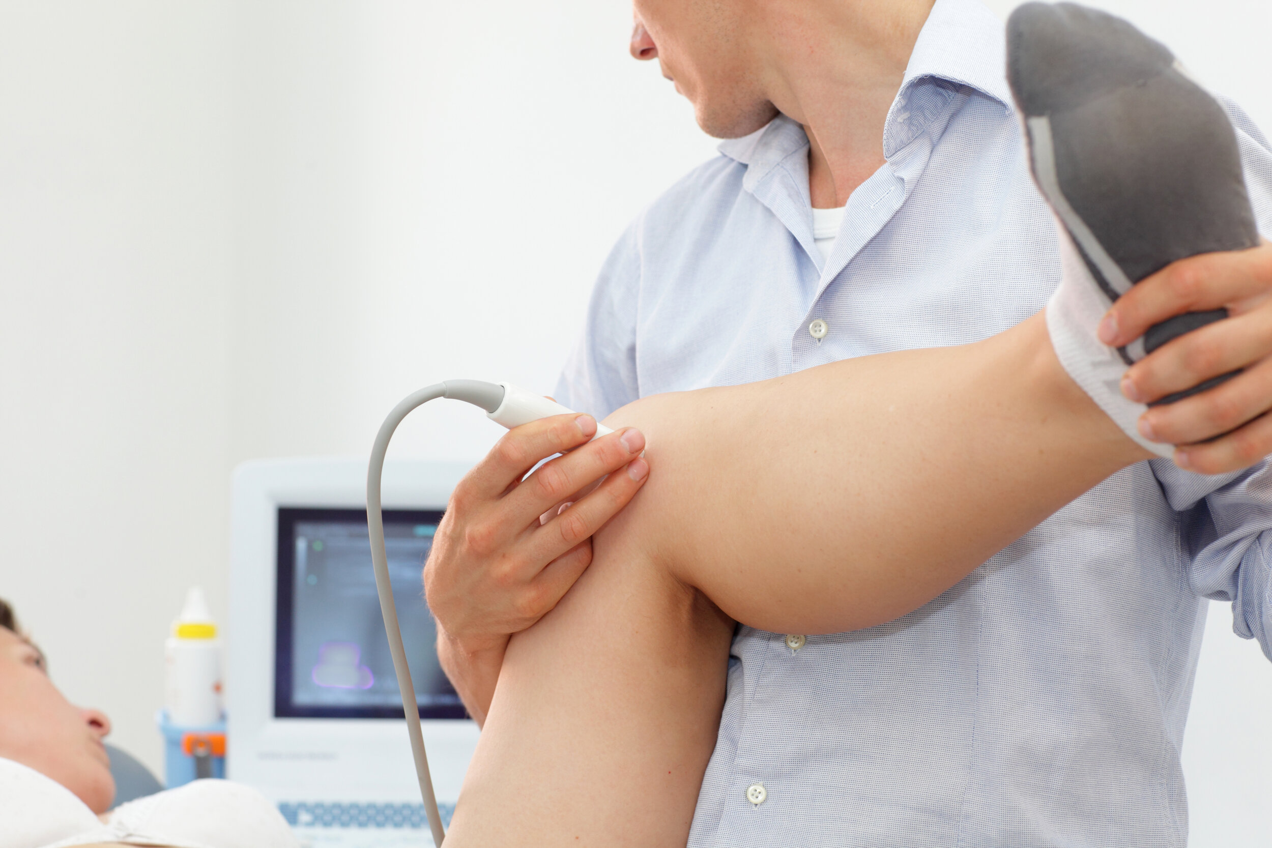
Ultrasound scanners consist of a computer console, video display screen and an attached transducer. The clinician applies a small amount of gel to the area under examination and places the transducer there. The gel allows sound waves to travel back and forth between the transducer and the area under examination. The ultrasound image is immediately visible on a video display screen that looks like a computer monitor. Once the imaging is complete, the clear ultrasound gel will be wiped off your skin. The ultrasound gel does not stain or discolour clothing. Depending on the area of examination, patient will be asked to sit or lie in suitable position. For some ultrasound exams, the patient is positioned lying face-up or face-down on an examination table. The clinician may ask you to move the extremity being examined or may move it for you to evaluate the anatomy and function of the joint, muscle, ligament or tendon.
What will I experience during and after the procedure?
Most ultrasound exams are painless, fast and easily tolerated. Musculoskeletal ultrasound examination is usually completed within 15 to 30 minutes but may occasionally take longer. When the exam is complete, you may be asked to dress and wait while the ultrasound images are reviewed. After an ultrasound examination, you should be able to resume your normal activities immediately.
Who interprets the results and how do I get them?
The Podiatry & Physiotherapy Clinics clinicians are trained in musculoskeletal ultrasound examination will interpret images exams. A clinician will analyse the images and correlate with musculoskeletal assessment and can provide diagnosis/report to patient and can also send a signed assessment report to the doctor. The clinicians can also discuss outcome with patient after the complete musculoskeletal assessment.
What are the benefits vs risks?
Benefits
Ultrasound is widely available, easy-to-use and less expensive than most other imaging methods.
Ultrasound imaging is extremely safe and does not use radiation.
Ultrasound scanning gives a clear picture of soft tissues that do not show up well on x-ray images.
Ultrasound provides real-time imaging, making it a good tool for guiding minimally invasive procedures such as ultrasound guided injections in joints and soft tissues as well as fluid aspiration.
Patients with cardiac pacemakers and certain types of metallic implants or fragments in the body cannot be safely exposed to the strong magnetic field required for magnetic resonance imaging (MRI); however, patients can safely receive ultrasound imaging.
Ultrasound is also an excellent alternative to MRI for claustrophobic patients.
Compared to MRI, ultrasound may provide greater internal detail when assessing soft tissue structures such as tendons and nerves.
Because ultrasound images are captured in real time, they can show the movement of a soft tissue structure such as a tendon, joint or an extremity.
Ultrasound imaging is faster than MRI and does not require the patient to remain completely still, allowing infants to be imaged without sedation.
Risks
Standard diagnostic ultrasound has no known harmful effects on humans.
What are the limitations of Ultrasound Imaging of the Musculoskeletal System?
Ultrasound has difficulty penetrating bone and, therefore, can only see the outer surface of bony structures and not what lies within. For visualizing internal structure of bones or certain joints, other imaging modalities such as MRI are typically used. There are also limitations to the depth that sound waves can penetrate; therefore, deeper structures in larger patients may not be seen easily.